

MOMENTUM™ Imager
Magnetic Particle Imaging system
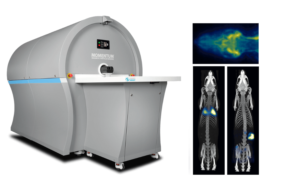
Left: MOMENTUM Imager. Upper right: Neurovascular perfusion and blood pool imaging. Middle: Stem cells within the lungs in a rat. Lower-right: Orthotopic breast tumor in a rat.
MPI(Magnetic Particle Imaging) 기술은 직접적으로 superparamagnetic nanoparticle tracer를 측정하는 이미징 기술로,
높은 sensitivity와 절대 정량이 가능한 이미징 기술로 시료의 깊이에 제한 없이 수개월에 이르기까지 모니터링이 가능합니다.
이러한 기술이 접목된 MOMENTUM™ Imager는cancer, infectious disease, cell tracking, vascular research 등의 분야에서
탁월한 이미징 데이터를 제공합니다.
- Longitudinal cell tracking
- Real-time vascular function
- Cancer & immunology
- Quantitative 2D & 3D imaging
Control every aspect of your experiment
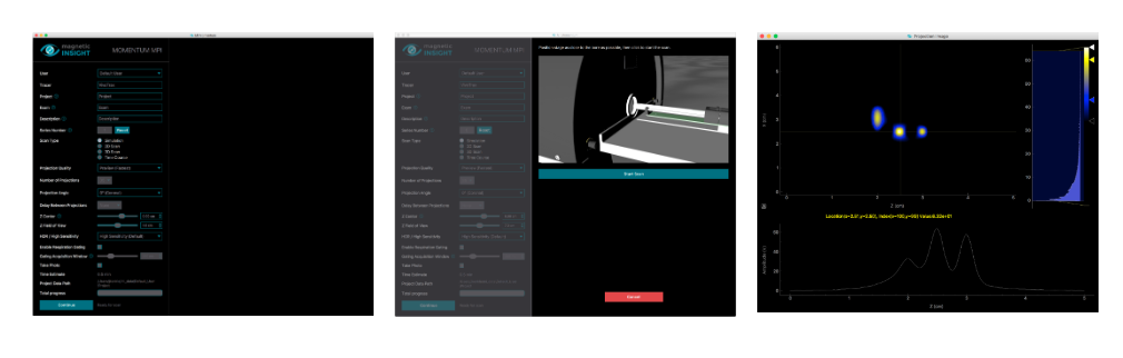
Straightforward acquisition settings One-click scanning and monitoring Quick image viewer
Magnetic Particle Imaging: A New Imaging Modality
MPI is the most promising emerging imaging technology in the last 20 years and is expected to change the landscape of modern medical imaging and in vivo translational research.”
– IWMPI 2014
Magnetic Particle Imaging (MPI) is a new imaging modality that directly detects iron oxide nanoparticle tracers using time-varying magnetic fields. Because the tracer is not normally found in the body, MPI images have exceptional contrast and high sensitivity.
Magnetic Particle Imaging uses a unique geometry of magnetics to create a field free region (FFR). This is something you may have experienced when pointing two magnets at each other. That sensitive point controls the direction of a nanoparticle.

Rapidly Moving the FFR causes a “flip” in the magnetic direction of an SPIO nanoparticle which induces a signal in a receive coil. Since we know where the sensitive point is at all times, we can assign the signal to the known position to produce a quantitative MPI image.

